Beranda
/ Anatomy Of Chest / The Respiratory System Anatomical Chart | Lung Anatomy Poster / 12 cm (5 in) in length, 8 cm (3.5 in) wide, and 6 cm (2.5 in) in thickness.
Anatomy Of Chest / The Respiratory System Anatomical Chart | Lung Anatomy Poster / 12 cm (5 in) in length, 8 cm (3.5 in) wide, and 6 cm (2.5 in) in thickness.
Insurance Gas/Electricity Loans Mortgage Attorney Lawyer Donate Conference Call Degree Credit Treatment Software Classes Recovery Trading Rehab Hosting Transfer Cord Blood Claim compensation mesothelioma mesothelioma attorney Houston car accident lawyer moreno valley can you sue a doctor for wrong diagnosis doctorate in security top online doctoral programs in business educational leadership doctoral programs online car accident doctor atlanta car accident doctor atlanta accident attorney rancho Cucamonga truck accident attorney san Antonio ONLINE BUSINESS DEGREE PROGRAMS ACCREDITED online accredited psychology degree masters degree in human resources online public administration masters degree online bitcoin merchant account bitcoin merchant services compare car insurance auto insurance troy mi seo explanation digital marketing degree floridaseo company fitness showrooms stamfordct how to work more efficiently seowordpress tips meaning of seo what is an seo what does an seo do what seo stands for best seotips google seo advice seo steps, The secure cloud-based platform for smart service delivery. Safelink is used by legal, professional and financial services to protect sensitive information, accelerate business processes and increase productivity. Use Safelink to collaborate securely with clients, colleagues and external parties. Safelink has a menu of workspace types with advanced features for dispute resolution, running deals and customised client portal creation. All data is encrypted (at rest and in transit and you retain your own encryption keys. Our titan security framework ensures your data is secure and you even have the option to choose your own data location from Channel Islands, London (UK), Dublin (EU), Australia.
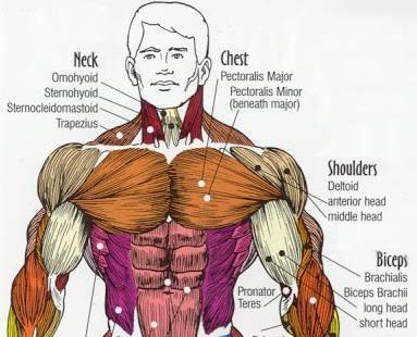
Anatomy Of Chest / The Respiratory System Anatomical Chart | Lung Anatomy Poster / 12 cm (5 in) in length, 8 cm (3.5 in) wide, and 6 cm (2.5 in) in thickness.. It is made up of the manubrium superiorly, the body and the xiphisternum (figure 1).the manubrium has an upper central depression, the suprasternal notch. Swensen fund for innovation in teaching. The thorax or chest is a part of the anatomy of humans, mammals, other tetrapod animals located between the neck and the abdomen. The chest anatomy includes the pectoralis major, pectoralis minor and the serratus anterior. Here, we break down the anatomy of your chest muscles.
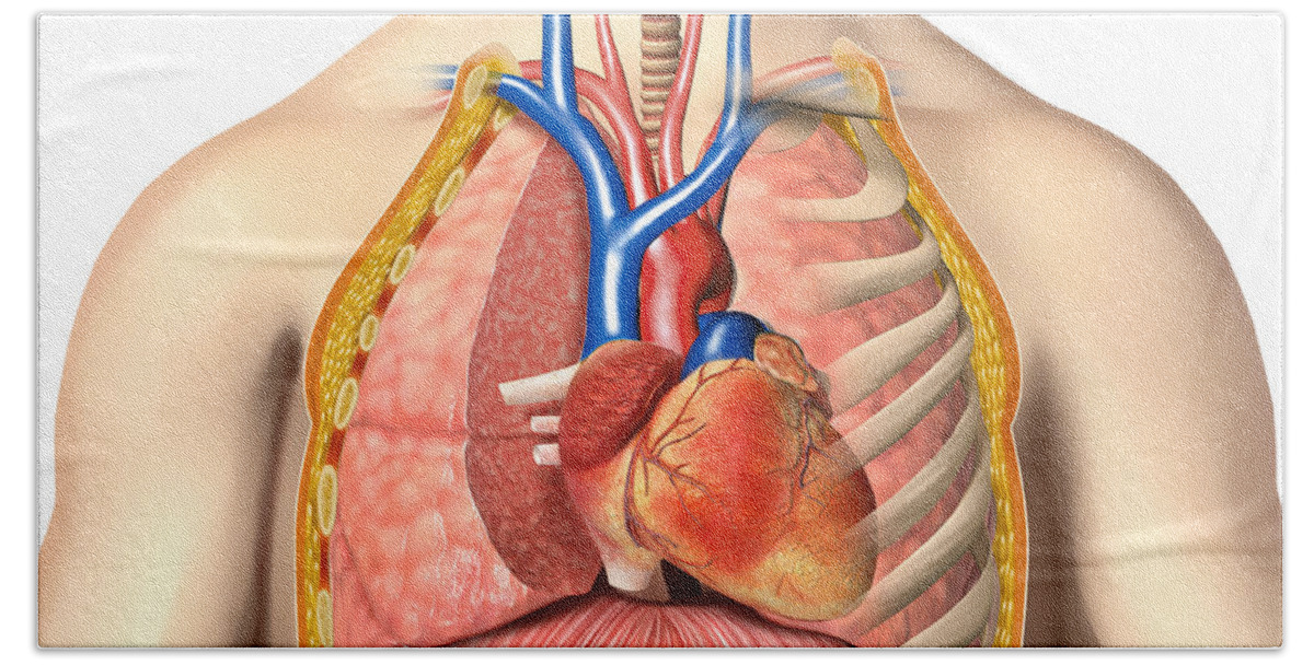
Organs & structures of the chest heart. The chest is the area of origin for many of the body's systems as it houses organs such as the heart, esophagus, trachea, lungs, and thoracic diaphragm. This atlas is a comprehensive and affordable learning tool for medical students and residents and especially for radiologists and pneumologists. Structures to identify • heart • lungs • mediastinum • pleural space • chest wall • …everything else! The dominant muscle in the upper chest is the pectoralis major.
Male Chest Anatomy Of Thorax Bath Towel for Sale by ... from render.fineartamerica.com The pec major) is the one that commands the most real estate. First i'll do an intro to the different organs and structures in the chest, and then i'll go over some images showing their locations. The chest is the area of origin for many of the body's systems as it houses organs such as the heart, esophagus, trachea, lungs, and thoracic diaphragm. The circulatory system does most of its work. This page provides an overview of the chest muscle group. It provides access to ct images in the axial plane, allowing the user to learn and review the lung anatomy interactively. Swensen fund for innovation in teaching. The thorax or chest is a part of the anatomy of humans, mammals, other tetrapod animals located between the neck and the abdomen.
Of the two chest muscles, the pectoralis major (a.k.a.
Thoracic cavity, also called chest cavity, the second largest hollow space of the body. The chest or thorax is the region between the neck and diaphragm that encloses organs, such as the heart, lungs, esophagus, trachea, and thoracic diaphragm. (1) the pectoralis major, and (2) the pectoralis minor. The circulatory system does most of its work. An overview of the anatomy visible in a transverse computed axial tomographical image of the thorax (and part of the abdomen) performed with intravenous cont. Anatomy of the thorax, heart, abdomen and pelvis recommended text gray's anatomy for students. Anatomy of right side chest pain. About the 6th week, the somites differentiate into the sclerotomes and the dermatomyotomes. However, the classical anatomical descriptions in textbooks make it difficult to gain full mastery of this subject, because the books usually deal with its elements separately. This atlas is a comprehensive and affordable learning tool for medical students and residents and especially for radiologists and pneumologists. It provides access to ct images in the axial plane, allowing the user to learn and review the lung anatomy interactively. The chest wall is comprised of skin, fat, muscles, and the thoracic skeleton. The human thorax includes the thoracic cavity and the thoracic wall.
However, the classical anatomical descriptions in textbooks make it difficult to gain full mastery of this subject, because the books usually deal with its elements separately. (1) the pectoralis major, and (2) the pectoralis minor. In insects, crustaceans, and the extinct trilobites, the thorax is one of the three main divisions of the creature's body, each of which is in turn composed of multiple segments. Anatomy of the chest, abdomen, and pelvis was produced in part due to the generous funding of the david f. It provides access to ct images in the axial plane, allowing the user to learn and review the lung anatomy interactively.
Targeting A Stubborn Chest - Working The Pecs! from www.bodybuilding.com About the 6th week, the somites differentiate into the sclerotomes and the dermatomyotomes. It is made up of the manubrium superiorly, the body and the xiphisternum (figure 1).the manubrium has an upper central depression, the suprasternal notch. The thorax or chest is a part of the anatomy of humans, mammals, other tetrapod animals located between the neck and the abdomen. Of the two chest muscles, the pectoralis major (a.k.a. Here's how science can help you grow! Table 1.1 lists the major anatomic structures within the thorax that are discussed. Anatomy of the chest and shoulder, anatomy of the chest organs, anatomy of the chest wall, anatomy of the chest wall and pleura, anatomy of upper chest area, human. This atlas is a comprehensive and affordable learning tool for medical students and residents and especially for radiologists and pneumologists.
The human thorax includes the thoracic cavity and the thoracic wall.
In insects, crustaceans, and the extinct trilobites, the thorax is one of the three main divisions of the creature's body, each of which is in turn composed of multiple segments. The chest or thorax region of the upper body has a number of important organs that reside within it that may present with chest pain if they become compromised in. How to view the anatomical labels. Anatomy of right side chest pain. (1) the pectoralis major, and (2) the pectoralis minor. It provides protection to vital organs (eg, heart and major vessels, lungs, liver) and provides stability for movement. Basic thoracic anatomy and physiology an understanding of thoracic imaging requires knowledge of the anatomy being imaged, as described in this chapter, as well as the imaging techniques applied to the thorax, covered in chapter 2. This atlas is a comprehensive and affordable learning tool for medical students and residents and especially for radiologists and pneumologists. The chest is the area of origin for many of the body's systems as it houses organs such as the heart, esophagus, trachea, lungs, and thoracic diaphragm. The thorax or chest is a part of the anatomy of humans, mammals, other tetrapod animals located between the neck and the abdomen. Computed tomography (ct) of the chest can detect pathology that may not show up on a conventional chest radiograph(1). However, the classical anatomical descriptions in textbooks make it difficult to gain full mastery of this subject, because the books usually deal with its elements separately. Skandalakis chest wall embryogenesis the muscles of the chest develop from the somites found in the mesoderm.
First i'll do an intro to the different organs and structures in the chest, and then i'll go over some images showing their locations. Swensen fund for innovation in teaching. The chest or thorax region of the upper body has a number of important organs that reside within it that may present with chest pain if they become compromised in. How to view the anatomical labels. The pec major) is the one that commands the most real estate.
Anatomy of the thorax (CT) from www.imaios.com The chest or thorax is the region between the neck and diaphragm that encloses organs, such as the heart, lungs, esophagus, trachea, and thoracic diaphragm. The anatomic illustrations are presented as… The circulatory system does most of its work. An overview of the anatomy visible in a transverse computed axial tomographical image of the thorax (and part of the abdomen) performed with intravenous cont. Anatomy of the thorax, heart, abdomen and pelvis recommended text gray's anatomy for students. The chest is made up primarily of two muscles: Anatomy of right side chest pain. Basic thoracic anatomy and physiology an understanding of thoracic imaging requires knowledge of the anatomy being imaged, as described in this chapter, as well as the imaging techniques applied to the thorax, covered in chapter 2.
The human thorax includes the thoracic cavity and the thoracic wall.
Structures to identify • heart • lungs • mediastinum • pleural space • chest wall • …everything else! Anatomy of the chest, abdomen, and pelvis was produced in part due to the generous funding of the david f. The chest wall is comprised of skin, fat, muscles, and the thoracic skeleton. Anatomy of the chest and shoulder, anatomy of the chest organs, anatomy of the chest wall, anatomy of the chest wall and pleura, anatomy of upper chest area, human. Organs & structures of the chest heart. First i'll do an intro to the different organs and structures in the chest, and then i'll go over some images showing their locations. Learn about each of these muscles, their locations, functional anatomy and exercises for them. Anatomy of the thorax, heart, abdomen and pelvis recommended text gray's anatomy for students. The pec major) is the one that commands the most real estate. Swensen fund for innovation in teaching. However, the classical anatomical descriptions in textbooks make it difficult to gain full mastery of this subject, because the books usually deal with its elements separately. The chest is made up primarily of two muscles: Table 1.1 lists the major anatomic structures within the thorax that are discussed.